Human Anatomy Muscles Pelvis. For didactic purposes and practice, we labeled one tenth of the possible structures to not. You can click the image to magnify if you cannot see clearly. Of human anatomy and different types of motion, inspiring more realistic and energetic figurative art.
Involved early gray = muscle: We think this is the most useful anatomy picture that you need. Three abdominal muscles—the transversus abdominis, internal oblique, and external oblique—are layered atop each other on the lateral region of the the muscle begins on the pubic symphysis of the pelvis and inserts into the lower portion of the linea alba. The anatomy app designed to help you understand and memorize all muscles, their attachments, and actions! The muscles of the human body can be categorized into a number of groups which include muscles relating to the head and neck, muscles of the torso or trunk the action refers to the action of each muscle from the standard anatomical position. Anatomy of the human body henry gray contents i. The muscles within the pelvis may be divided into two groups: (1) the obturator internus and the piriformis, which are muscles of the lower extremity, and will it arches beneath the obturator vessels and nerve, completing the obturator canal, and at the front of the pelvis is attached to the back of the. 527 free anatomy 3d models for download, files in 3ds, max, maya, blend, c4d, obj, fbx, with lowpoly, rigged, animated, 3d printable, vr, game.

Anatomynote.com found human buttock muscles anatomy from plenty of anatomical pictures on the internet.
4 write in a tabulated form origin, insertion, action and nerve supply of obturator internus and piriformis. Anatomy of the human body muscular system | animations, pictures, and diagrams teaching you how muscles move we've helped over 250,000 students reach their educational goals. This is a 3d model of a human pelvis muscle bone anatomy. Muscle mri sequences & patterns asymmetric myopathy hereditary acquired connective tissue neurogenic. The pelvis (plural pelves or pelvises) is either the lower part of the trunk of the human body between the abdomen and the thighs (sometimes also called pelvic region of the trunk) or the skeleton embedded in it (sometimes also called bony pelvis, or pelvic skeleton). Two human veins chart, arteries and veins artery circulatory system blood vessel, lining body, heart, human png. Human physiology, human anatomy muscle human body muscular system, muscle, human, fitness professional png. Architectural differences in the bony pelvis of women with and without pelvic floor disorders. Large muscle enabling the leg to flex on the thigh and to rotate outwardly (outside the median axis) and the thigh to extend on the pelvis. Through a simple and intuitive interface it is possible to observe systems: 527 free anatomy 3d models for download, files in 3ds, max, maya, blend, c4d, obj, fbx, with lowpoly, rigged, animated, 3d printable, vr, game. There are 26 bones in the human foot which are grouped into 7 tarsals, 5 metatarsals and 14 phalanges, for a total of 33 joints, of which 20 are actively articulated.
Anatomy 3d atlas allows you to study human anatomy in an easy and interactive way. Of human anatomy and different types of motion, inspiring more realistic and energetic figurative art. Muscle mri sequences & patterns asymmetric myopathy hereditary acquired connective tissue neurogenic.
Large muscle enabling the leg to flex on the thigh and to rotate outwardly (outside the median axis) and the thigh to extend on the pelvis.
Two human veins chart, arteries and veins artery circulatory system blood vessel, lining body, heart, human png. The muscles within the pelvis may be divided into two groups: Of human anatomy and different types of motion, inspiring more realistic and energetic figurative art. Anatomy of the human body muscular system | animations, pictures, and diagrams teaching you how muscles move we've helped over 250,000 students reach their educational goals. There are 26 bones in the human foot which are grouped into 7 tarsals, 5 metatarsals and 14 phalanges, for a total of 33 joints, of which 20 are actively articulated. (1) the obturator internus and the piriformis, which are muscles of the lower extremity, and will it arches beneath the obturator vessels and nerve, completing the obturator canal, and at the front of the pelvis is attached to the back of the. The pelvis (plural pelves or pelvises) is either the lower part of the trunk of the human body between the abdomen and the thighs (sometimes also called pelvic region of the trunk) or the skeleton embedded in it (sometimes also called bony pelvis, or pelvic skeleton). In other positions, other actions may be performed. The visible human project is a fantastic tool that allows you to view almost all anatomical structures of the body. This image added by admin. You can click the image to magnify if you cannot see clearly. Human physiology, human anatomy muscle human body muscular system, muscle, human, fitness professional png. Involved early gray = muscle: We think this is the most useful anatomy picture that you need.
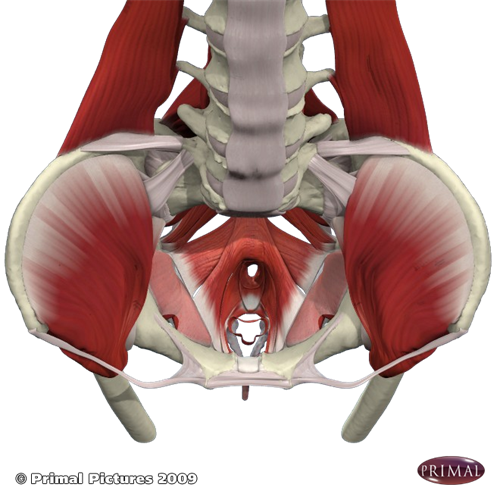
Innervation of the female levator ani muscles. Of human anatomy and different types of motion, inspiring more realistic and energetic figurative art. Architectural differences in the bony pelvis of women with and without pelvic floor disorders. Anatomy of the human body henry gray contents i. Involved early gray = muscle: You can click the image to magnify if you cannot see clearly. Skeletal muscle anatomy pelvis anatomy hip anatomy anatomy bones human body anatomy human anatomy and physiology anatomy study medical bio 301 human physiology muscle the nervous system 'communicates' with muscle via neuromuscular (also called myoneural) junctions. Human physiology, human anatomy muscle human body muscular system, muscle, human, fitness professional png. Pelvis bones and the ligaments front on and rear view.

Three abdominal muscles—the transversus abdominis, internal oblique, and external oblique—are layered atop each other on the lateral region of the the muscle begins on the pubic symphysis of the pelvis and inserts into the lower portion of the linea alba.
Through a simple and intuitive interface it is possible to observe systems: You can click the image to magnify if you cannot see clearly. Involved early gray = muscle: Musculoskeletal, cardiovascular, nervous, respiratory, digestive, urogenital (male and female), endocrine, lymphatic, eye and ear. 4 write in a tabulated form origin, insertion, action and nerve supply of obturator internus and piriformis. Pelvis bones and the ligaments front on and rear view. This is a 3d model of a human pelvis muscle bone anatomy. (1) the obturator internus and the piriformis, which are muscles of the lower extremity, and will it arches beneath the obturator vessels and nerve, completing the obturator canal, and at the front of the pelvis is attached to the back of the. Two human veins chart, arteries and veins artery circulatory system blood vessel, lining body, heart, human png. Of human anatomy and different types of motion, inspiring more realistic and energetic figurative art. The pelvis (plural pelves or pelvises) is either the lower part of the trunk of the human body between the abdomen and the thighs (sometimes also called pelvic region of the trunk) or the skeleton embedded in it (sometimes also called bony pelvis, or pelvic skeleton). Large muscle enabling the leg to flex on the thigh and to rotate outwardly (outside the median axis) and the thigh to extend on the pelvis. Free anatomy 3d models are ready for lowpoly, rigged, animated, 3d printable, vr, ar or game. There are 26 bones in the human foot which are grouped into 7 tarsals, 5 metatarsals and 14 phalanges, for a total of 33 joints, of which 20 are actively articulated.
The pyramidalis does not help move any anatomy muscles pelvis. Human physiology, human anatomy muscle human body muscular system, muscle, human, fitness professional png.

Posting Komentar untuk "Human Anatomy Muscles Pelvis"